
Emphysema: What It Is and How It Differs from COPD
Emphysema is the pattern of COPD where the air sac walls are permanently damaged. What the tests show, what a scan line means and what care can change.
 Written byDr. Mohammad Fawzi Katranji
Published

Medical review pending. This page is written for review by consultant pulmonologist Dr. Mohammad Fawzi Katranji, whose four American Board certifications cover Internal Medicine, Pulmonary Medicine, Critical Care Medicine and Sleep Medicine. A review date is added once that review is complete.
You have been handed a CT report with the word emphysema on it, or a consultant has said there is "a bit of emphysema" in your lungs. It means permanent damage to the walls of the air sacs, and it is a pattern inside COPD rather than a separate disease.
The damage does not grow back, and no inhaler, tablet or supplement repairs it. Some of the picture can still change: how much the pattern affects your breathing, how fast it moves, and what happens to the exposures still in play.
People arrive at this from different directions. One has a cough that will not clear after a winter of chest infections, another notices the stairs at work have become harder than they were, and a third went into a scanner for something else and came out with a line about emphysema. Each route leads to the same questions about damage, airflow and what happens next.
Knowing which pattern is on your report, and knowing your spirometry numbers, makes the next appointment more useful.
Key takeaways
- Emphysema means damage to the walls of the air sacs (alveoli), and it sits inside COPD alongside chronic bronchitis.
- The confirming test is spirometry once a bronchodilator has been given: an FEV1/FVC ratio below 0.7.
- Emphysema that shows on a CT ordered for something else needs reviewing with spirometry, and on its own it raises lung cancer risk.
- For anyone with COPD who smokes, stopping comes ahead of every other treatment.
What is emphysema?
Emphysema is permanent damage to the air sacs (alveoli) at the tips of the airways. The walls between them break down, the tissue loses its stretchiness, and air moves in more easily than it comes out. Nothing restores the walls that have been lost.
The lungs hold more than 300 million of these sacs, according to the American Thoracic Society's patient series. Each one is a dead end, and oxygen crosses into the blood through the thin wall it shares with its neighbour, while carbon dioxide crosses the other way. That exchange is what the words gas exchange mean, and it is the job a damaged wall does badly.
In emphysema the walls break down and neighbouring sacs merge into larger, flatter spaces, so there is less surface for that exchange to happen across. The society's own wording is that the walls "are damaged and lose their stretchiness".
The change is permanent. Treatment can ease breathlessness, protect what is left and lower the risk of a flare-up; it cannot rebuild tissue that has gone. If a scan report has left you with questions, book an appointment and bring the report with you.
What the damage does to your breathing
Air goes in and does not come all the way out. Sacs that no longer empty trap air, the chest sits fuller than it should, and the lungs are described as hyperinflated, as the American Thoracic Society explains.
Breathlessness on stairs tends to come first. A cough that hangs on through the winter, sputum most mornings and a wheeze on cold nights are the other usual presentations.
NICE lists the symptoms that make COPD worth suspecting once someone is past 35 with a smoking history. It starts with breathlessness on effort and a cough that will not settle. Sputum on most days, bronchitis that returns each winter and a wheeze on cold nights belong to the same picture.
The NICE guideline NG115 also asks about weight loss, tiring on exertion and breathlessness that wakes you at night. Swollen ankles and fatigue belong on that list, along with chest pain and coughing up blood, though both of those are uncommon in COPD and point toward another diagnosis.
The Medical Research Council scale grades breathlessness by what brings it on. Grade 1 means trouble only on strenuous exercise; at grade 5, leaving the house is too much, or dressing leaves you short of breath. Your grade says more about your week than any spirometry number.
None of these symptoms proves emphysema. The same breathlessness can come from the heart, from anaemia or from being out of condition, which is why a first assessment includes a chest X-ray and a blood count next to the breathing tests. Breathlessness by itself has a long list of possible causes, and the pattern you describe helps narrow it.
Is emphysema a type of COPD?
The American Thoracic Society describes COPD as chronic bronchitis, emphysema, or both at once in the same person. Emphysema is one pattern inside that umbrella, and the label alone says little about how much airflow is left.
Which pattern dominates changes what the assessment looks for. Emphysema turns the questions toward the air sacs, gas exchange and hyperinflation; chronic bronchitis turns them toward the airways and sputum. Most people carry some of each, and the imaging and spirometry results decide how a letter describes it.
That is why a report naming emphysema does not measure your COPD. The airflow numbers do, and they come from spirometry. The whole-disease view sits in COPD: the whole-disease view.
How emphysema differs from chronic bronchitis and asthma
Chronic bronchitis and emphysema describe different parts of the same lung. In the first, inflamed airways produce a cough with sputum. In the second, the damage sits in the air sacs. Many people have both.
Asthma sits outside that definition. Variability splits the two in clinic: obstruction in COPD holds and progresses, while in asthma it shifts from day to day and week to week.
NICE sets out which history features help. A smoking past is near-universal in COPD and only possible in asthma. Symptoms beginning before 35 are rare in COPD and frequent in asthma. A daily productive cough is usual in COPD and unusual in asthma, and breathlessness that persists and progresses belongs to COPD where asthma's comes and goes.
Waking in the night with breathlessness or wheeze counts the other way, as does a symptom pattern that swings from one day to the next. Peak flow logged twice a day for a couple of weeks shows that swing, which is why it is used when the picture is unclear.
Some people have both conditions, and the guideline expects a mixed history to be recognised as such. COPD and asthma compared takes that overlap further.
What causes it, and why non-smokers get it
Tobacco smoke is the commonest exposure behind COPD, and it arrives in several forms: cigarettes, cigars and waterpipe (shisha) belong to the same exposure class. The guideline builds its suspicion on a smoking history plus symptoms, once someone is past 35.
The appointment should also cover the dust and fumes you meet at work. That same NICE guideline asks about occupational hazards: construction dust, mining, welding, paint, cleaning chemicals and vehicle exhaust all count. Smoke from wood or biomass burned indoors, and heavy air pollution, are the other exposures that come up.
Some people with none of those exposures still develop emphysema. The inherited cause that gets tested is alpha-1 antitrypsin deficiency, a protein problem that leaves lung tissue unprotected. NICE asks for that blood test when the condition appears early, when someone has smoked little or never, or when relatives have had it, and it also asks about lung or liver disease in the family or in the person's own past.
One narrow situation is worth knowing. When a scan shows emphysema in someone who has never smoked, with normal spirometry and no symptoms, the guidance asks the doctor to look for those alternatives and then to reassure the person that the finding is unlikely to get worse. Ask which of those situations fits you.
How it is assessed: spirometry and the bronchodilator
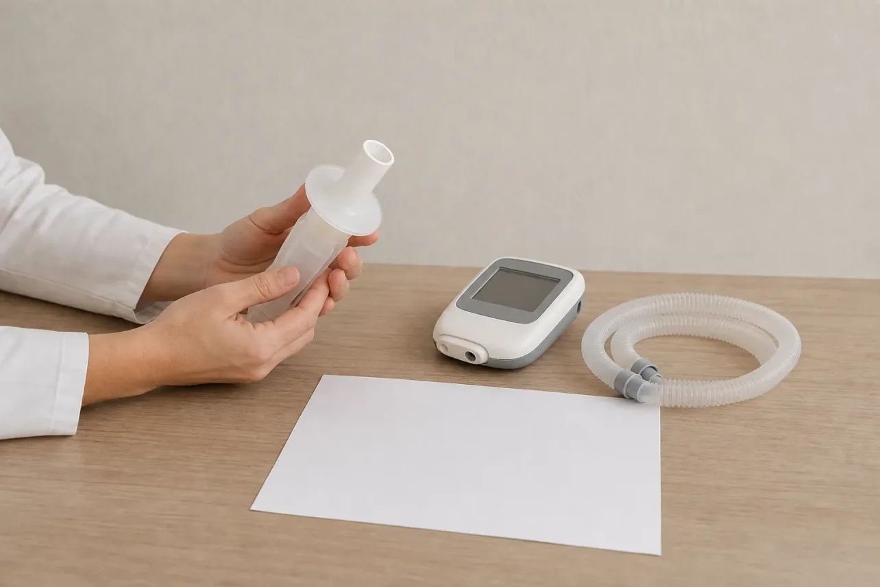
Spirometry is the test that settles the question. You blow into a mouthpiece with everything you have, for as long as you can, and then repeat it after a bronchodilator inhaler has been given. The machine reports how much you blew out in total (FVC) and how much came out in the first second (FEV1), and the ratio between them matters most.
NICE confirms COPD when the ratio of FEV1 to FVC stays at 0.7 or lower once the bronchodilator has been given. The American Thoracic Society goes further, putting spirometry first among the tests that decide whether someone has COPD. How a spirometry test is done walks through the appointment itself.
The ratio says whether obstruction is there. The FEV1 percentage of predicted says how much. NICE's table 4 carries more than one labelling scheme, so two reports can describe the same FEV1 with different words:
| FEV1 (% of predicted) after the bronchodilator | Grade usually written |
|---|---|
| 80% or more | Mild |
| 50% to 79% | Moderate |
| 30% to 49% | Severe |
| Under 30% | Stage 4 in the schemes NICE reproduces |
An older person whose ratio sits below 0.7 but who has none of the typical symptoms needs other diagnoses considered too. A younger person with the right symptoms can still have COPD with a ratio above 0.7. One reversibility test can mislead unless the change in FEV1 is large, so the whole picture decides.
When spirometry leaves something open, or a report already names emphysema, pulmonary function tests add lung volumes and a gas transfer measurement (TLCO). CT comes in when symptoms look worse than the spirometry numbers suggest, when another lung disease is possible, or as part of an assessment for lung volume reduction.
What the words on your scan report mean
A radiologist's line describes where the damage sits and what it looks like. It does not grade your breathlessness, and it does not by itself tell you how much airflow you have lost.
Mild. A word about amount, used in imaging reports and in spirometry grading alike. Mild looking changes on a scan and mild airflow obstruction are two separate measurements, so ask for your ratio and FEV1 as well.
Centrilobular. The damage starts at the centre of each small lung unit, around the fine airways, and this is the pattern most often linked with smoking.
Paraseptal. The damage runs along the outer edges of the lung, just under the pleura, and it can form larger air-filled spaces there.
Bullous. From bulla, a single air-filled space that has replaced several sacs. A bulla takes no part in gas exchange, and a large one can press on the lung beside it.
NICE asks for the finding to be reviewed by a respiratory clinician, with spirometry, notes that emphysema on CT counts as a risk factor for lung cancer in its own right, and, in a non-smoker, asks about relatives with lung or liver disease before testing for alpha-1 antitrypsin deficiency.
At the appointment, ask which pattern the report describes, what your FEV1/FVC ratio and FEV1 came out at, and whether anything in your history changes what should be tested next.
What care aims at
Care aims at the airflow obstruction, the symptoms it causes and the exposures still doing damage. For anyone who smokes, stopping is the step the American Thoracic Society puts ahead of all others. That holds even when spirometry is normal and emphysema showed up only on a CT scan, where NICE still offers cessation support and a warning about higher risk.
Inhaled treatment can ease breathlessness for some people. The guideline expects the diagnosis to be looked at again when someone reports a marked improvement with an inhaler, because that pattern fits asthma better.
Pulmonary rehabilitation combines supervised exercise with education about breathing, and it is offered as part of COPD care. Vaccination belongs in the plan as well: a flu jab each year, plus pneumonia and COVID-19 vaccines when they are recommended.
Oxygen is not a general treatment for breathlessness. Long-term oxygen is considered only where blood oxygen levels meet a defined threshold, and some people are referred for assessment for lung volume reduction.
Breathlessness that arrives suddenly, or that is severe while sitting still, is an emergency: call 998. For slower changes, COPD flare-up warning signs sets out what to watch for. A scan report or a spirometry printout you cannot interpret can be read alongside your history: book an appointment and bring both.
Questions people ask about emphysema
What is severe emphysema?
Severe describes how much the airflow is reduced on spirometry, not how much damage shows on a scan. In the grading NICE reproduces it means an FEV1 between 30% and 49% of predicted after a bronchodilator, with an FEV1/FVC ratio under 0.7. Your own report holds your figures.
What are some home remedies for emphysema?
Nothing you do at home repairs damaged air sacs. Stopping smoking if you smoke, keeping active, staying up to date with flu and pneumonia vaccines, and completing pulmonary rehabilitation if it is offered all help. New or worse breathlessness needs assessment.
Does COPD make you tired and weak?
Tiredness is a symptom NICE asks about, and so are tiring on exertion and broken sleep from breathlessness. It can have more than one cause, including poor sleep, the effort breathing takes, and a separate problem such as sleep apnea. Describe the pattern at your appointment.
References
Chronic obstructive pulmonary disease in over 16s: diagnosis and management (NG115). NICE guideline, published 5 December 2018, last updated 26 July 2019.
COPD patient resources. American Thoracic Society.
Medical information. This article is educational and does not replace individual medical advice. In an emergency call 998.